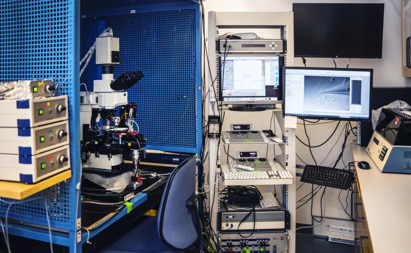
B10 Platform
Electrophysiology Core Facility
The B10 set-up consists of a HEKA EPC10 double-patch clamp amplifier, an Olympus BX51WI microscope with multi-channel fluorescence capability and differential interference contrast for live imaging and optical manipulation of cells (e.g. optogenetics and calcium imaging), and a Hamamatsu C11440-10C camera. This set-up is also equipped with a Multielectrode Array (MEA) system and there is a possibility to perform micromanipulator assisted collection of single cells for qPCR (Patch-Seq). Located on B10, inside the cell culture laboratory, this set-up is ideal for, but not restricted to, investigation of in vitro generated and expanded cell lines and cultures.
Major applications this platform offers:
Patch clamp recording in vitro (cell cultures) and, to some extent, ex vivo (tissue preparations) to study intrinsic electrical properties of single cells, such as resting membrane potential, membrane resistance, action potential parameters, presence and function of various channels and receptors as well as synaptic connectivity, and to identify, characterize and quantify synaptic transmission. Our main focus is neural lineage (e.g. neurons and astrocytes), but other cell types can also be recorded (e.g. cardiomyocytes, β cells).
Extracellular field potential recordings, spontaneous or evoked, to study general network activity, or synaptic plasticity ex vivo in brain tissue preparations (predominantly in hippocampus – LTP, LTD).
Optogenetic manipulation of cell function to study, e.g. synaptic profile of isolated populations of the cells in cell cultures and tissue preparations.
Optical imaging of live cells in cultures and tissue preparations to monitor intracellular Ca2+ fluctuations and to identify population of interest.
Multielectrode Array (MEA) recordings of extracellular field potentials from cell cultures and tissue preparations.
Book Our Services:
For bookings and consultations, please contact:
Electrophysiology [at] med [dot] lu [dot] se (Electrophysiology[at]med[dot]lu[dot]se)


