
Researchers from all disciplines and experience levels at Lund Stem Cell Center participated in the competition, sharing the hidden beauty discovered within their work. From intricate scientific images to cozy lab environments, the submitted visual narratives, accompanied by creative titles and scientific stories, painted a vivid picture of the remarkable research conducted at the Center.
"The Image Competition represents an opportunity for researchers to communicate their work to a different and wider audience," explains Johan Jakobsson, Director of the Lund Stem Cell Center. "As we celebrate our 20th anniversary, we are thrilled to unveil the extraordinary beauty and knowledge discovered within our walls."
This year, the competition enlisted the expertise of Debra Voges, a curator from Lunds Konsthall, who carefully reviewed each submission. The evaluation process hinged on three essential criteria: visual quality, scientific story, and representation, ensuring a comprehensive assessment of each image.
With great excitement, the Lund Stem Cell Center proudly announces the winners of the Image Competition 2023, extending congratulations to their achievements. Alongside these entries, we express sincere appreciation to all participants who shared their scientific artistry with us.
In the spirit of discovery, innovation, and artistic expression, we now invite you to embark on a visual, scientific journey as we present these images for the first time. Explore the gallery below, where each image offers a unique glimpse into the captivating world of stem cell research, bridging the gap between art and science.
Explore our 2023 Image Gallery:
First Place:

Researcher: Janko Kajtez, Associate Researcher, Kirkeby Research Group*
Description: Mini brain tissue, commonly referred to as brain organoid, grown in a lab from human stem cells displays numerous bundles of long neuronal processes that emanate from tissue core. Brain organoids mimic the developing human brain in a laboratory dish and are used to study neurodegenerative disorders such as Parkinson’s disease.
Assessment: Beautiful, powerful, and intriguing image that highlights an area of relevant current scientific research. Accompanied by a concise and accessible story, it makes us curious about an aspect of science that we might not be familiar with but that could benefit many of us in the future.
*This image was captured during Janko Kajtez's tenure as a researcher in the Parmar Research Group.
Second Place:

Researcher: Sofie Mohlin, Principal Investigator, Mohlin Research Group
Description: This hen provides us with the eggs we use to study chick embryos to understand childhood cancer formation during fetal development.
Assessment: Refreshing and original take on the task, creative and successful use of humor to tell a story that has the potential to be controversial and highlights lesser-known aspects of scientific research.
Third Place:

Researcher: Alessandro Fiorenzano, Associate Researcher, and Edoardo Sossi, PhD student, Parmar Research Group
Description: Immunofluorescence staining of human embryonic stem cell derived ventral midbrain progenitors used for cell replacement therapy for Parkinson’s Disease. Some of the cells that will become dopaminergic neurons upon transplantation, replacing the ones lost in Parkinson’s Disease.
Assessment: Immaculately balanced composition, stunning colors and a great story worth sharing.

Researcher: Gustav Christensson, PhD student, Hammarlund Research Group
Description: Metastasis is the last step of tumor development. This microscope photograph shows sections of human tissues containing breast and lung cancer cells (in white) establishing metastases in brain tissue. The colored cells are white blood cells of different types, but are they fighting the cancer – or perhaps supporting it?

Researcher: Dimitra Zacharaki, associate researcher, Bourgine Research Group
Description: Neuroblastoma is a childhood cancer with poor prognosis that often metastasizes to bone. NeurOssicle is a human mini-bone organoid consisting of neuroblastoma cancer cells (yellow) and a human microenvironment (magenta) that developed under the skin of a mouse.

Researcher: Nicholas Bèchet, Postdoctoral researcher, Lindstedt Research Group
Description: Despite the fears they elicit in some people, spiders are in fact often culturally portrayed creators, and associated with wisdom, patience, and persistence. In the composition of this image, I was taken back to a text I once read which described a spider as the creator of the university, its web the invisible fabric of our reality.

Researcher: Nicholas Bèchet, Postdoctoral researcher, Lindstedt Research Group
Description: A quote from my favorite childhood film which I feel is embodied by this image, and a view shared in many romanticized myths. Imagine if you will here, a small fragment of the university planets, stars, asteroids, wormholes, and comets all connected by the Webs of Creation.

Researcher: Nicholas Bèchet, Postdoctoral researcher, Lindstedt Research Group
Description: All things have a beginning and an end, the same is true of stars, only instead, of slowly fading from existence they release fire and brimstone, reducing themselves, and all the life they once supported to oblivion. Perhaps the greater kindness.

Researcher: Sofie Mohlin, Principal Investigator, Mohlin Research Group
Description: The heart of a chick embryo continues to beat hours after removal from the rest of the embryonic body. Intact aorta and chambers in this truly heart-shaped organ.
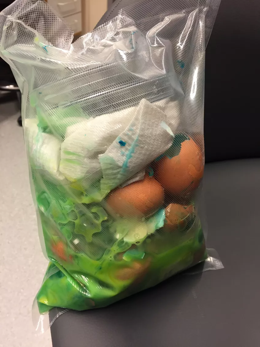
Researcher: Sofie Mohlin, Principal Investigator, Mohlin Research Group
Description: The waste produced from experiments addressing how childhood cancer forms during fetal development, using fertilised eggs and chick embryos.

Researcher: Johanna Farley, research assistant, and Madeleine Schwalbe, bachelor’s student, Gopal Research Group
Description: Even when you don’t get the results you expect, you can always make beautiful art out of it. This is composed using fluorescent images of C. elegans germlines which have miraculously formed into letters of the alphabet!

Researcher: Johanna Farley, research assistant, and Madeleine Schwalbe, bachelor’s student, Gopal Research Group
Description: Did you also miss the Northern Lights in Lund? This image depicts C. elegans (a microscopic worm) labelled with green fluorescent protein in the extracellular matrix. Equally as beautiful, the microscopic equivalent to the great natural phenomenon of the aurora borealis!

Researcher: Josefin Johansson, master’s student, Leigh Research Group
Description: The skull of a salamander larva with snout downwards, laying on its back. The collagen inside the cartilage is labelled with green fluorescent protein (GFP) and the surrounding tissue is made transparent through tissue clearing. The labelling of collagen is used as a positive control for this experiment.

Researcher: Anna Fossum, Project Manager, FACS Core Facility
Description: Working with the FACS Facility means you need to have a lot of different skills! Sometimes you need to take things apart and sometimes you need to put things together.
Note: Lund Stem Cell Center gratefully acknowledges Debra Voges from Lunds Konsthall for her meticulous evaluation of this year's submissions.


