UniStem workshops 2020
1. Crispr-Cas9 – a revolutionary tool for gene therapy in human disease?
This workshop will start with an overview of the CRISPR technology, its novelty and applications. We will then examine the DNA sequence of a gene and look for potential cut sites, design guideRNAs and talk about how to perform an experiment and what happens in the DNA after the CRISPR-cut. This will be followed by an exercise in experimental design. We will then end by with a small discussion of the potential of CRISPR-technology in human gene-editing and the ethical considerations surrounding such an approach.
Contact
Pia Johansson, Platform manager CRISPR facility
Diahann Atacho, Postdoctoral researcher at Molecular Neurogenetics
2. Exploring the cell diversity of the brain
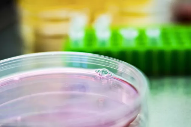
Did you know that neurons are not the only cell type in the human brain? We will tell you about all the cells that form the brain – including stem cells - and how these cells look like under the microscope. Thereafter, you will also have the opportunity to take care of them.
Contact
Ana Gonzalez Ramos, PhD student at Experimental Epilepsy Group
Eliška Waloschková, PhD student at Experimental Epilepsy Group
3. Engineering of human skeletal tissue in bioreactors
This workshop will first introduce the notion of 3D culture systems, as opposed to standard 2D approach traditionally used to grow cells in laboratories. We will explain how scaffolding material can be combined with bioreactor device; you will in fact have the opportunity to assemble your own system capable of hosting human cells. We will discuss how such 3D approach can be exploited in order to engineer human cartilage or human bone grafts, which could ultimately be implanted in patients needed tissue repair. Concrete examples of human engineered tissues will also be shown.
Contact
Paul Bourgine, Group leader at Molecular Skeletal Biology
Alejandro Garcia, Postdoctoral researcher at Molecular Skeletal Biology
4. Behavioral assessment of neural stem cell transplants – From animal models to functional assessment
In this workshop we will present how researchers assess neural stem cells in vivo. We will give an introduction about research on laboratory animals, how we create models of neurodegenerative diseases, and how to assess the neural stem cells in vivo. The focus is on Parkinson’s disease and the transplantation of cells into the brain of rodents. After the workshop the students will have a basic idea about the animal models used for transplantation, how to assess graft survival and the immune-response, and also how a neural stem cell transplant can improve the behavior of a rodent that has been rendered parkinsonian. The workshop will be based on presentations of the most commonly used behavioral test as well as some equipment. Due to the restricted nature of animal testing the practical part of the session will be limited.
Contact
Shelby Shrigley, PhD student at Developmental and Regenerative Neurobiology
Michael Sparrenius, Technician at Developmental and Regenerative Neurobiology
5. Snitta hjärnan och hitta transplantat
Ni kommer att få se hur man snittar en hjärna och hur man sen märker in den med speciella antikroppar för att kunna visualisera transplanterade stamceller. I mikroskopet letar vi sen upp cellerna och tittar på hur de transplanterade cellerna ser ut. Vi kommer även att visa hur man använder avancerad 3D imaging.
Contact
Ulla Jarl, Biomedicinsk analytiker i gruppen Developmental and Regenerative Neurobiology
Bengt Mattson, Developmental and Regenerative Neurobiology
Deirdre Hoban, Postdoctoral researcher at Developmental and Regenerative Neurobiology
6. Bioengineering lungs
For many patients with lethal lung diseases, lung transplantation is the only option to save their lives. However, there are not enough donor lungs to transplant all the patients that need a new lung. Thus, many scientists are looking into making new lungs in the lab. In our lab, we are studying which type of materials can be used to bioengineer lungs. The lung is made out of cells and many proteins that support the cells and tells them how to behave. Therefore, we want to build the lungs using the same proteins the lungs have. To do this we remove the cells and preserve the proteins of the lungs using a method called decellularisation. In this workshop you will learn more about decellularisation of lungs and will be able to perform the technique yourself on mouse and pig lungs.
Contact
Martina De Santis, PhD student at Lung Bioengineering and Regeneration
Hani Alsafadi, PhD student at Lung Bioengineering and Regeneration
7. Can you grow a brain in a dish?
The inaccessibility of functional human brain tissue and the inability of two-dimensional in vitro cultures to recapitulate the complexity and function of dopaminergic circuitries have made the study of human brain functions and dysfunctions challenging. In this workshop, we will show a method for differentiating human pluripotent stem cells into three-dimensional (3D) dopaminergic organoids, which mimic features of human ventral midbrain (VM) development by recreating authentic and functional dopamine neurons. The students will be able to see the organoid maturation at microscope over the time and perform medium change. After that, they will have the chance to watch short movies of immunolabelling-enabled 3D imaging of whole organoids in which we reconstruct regional identities, spatial organization and connectivity maps providing them an anatomical prospective of dopaminergic organoids.
Contact
Alessandro Fiorenzano, Postdoctoral researcher at Developmental and Regenerative Neurobiology
Jessica Giacomoni, PhD student at Developmental and Regenerative Neurobiology
Fredrik Nilsson, PhD student at Developmental and Regenerative Neurobiology
Edoardo Sozzi, Erasmus student at Developmental and Regenerative Neurobiology
8. Isolate and image cells: Flow cytometry, cell sorting and automated image analysis
This workshop will give students an introduction to the Fluorescence Activated Cell Sorting (FACS) technique. FACS enables the isolation of specific cell types such as rare stem cells based on the labeling of cellular structures with fluorescent molecules that emit light of different colors. The aim of sorting cells is to obtain pure populations of the cell types of interest for the study of e.g. gene and protein expression, cell development, cell shape, and intracellular processes. We will also demonstrate Cellomics High Content Screening and show automated image analysis of cells labeled with fluorescence. Applying automated image analysis makes it possible to rapidly analyze thousands of microscope images to e.g. measure protein expression, evaluate co-localization and distribution of targets in a cell, count and measure branching of neurites. (20 min)
Contact
Anna Hammarberg, MultiPark - Cellomics and Flow Cytometry Core Facility
Karolina Pircs, Researcher at Molecular Neurogenetics
9. Electrophysiology: Measuring electrical activity of neuronal cells
We will begin with a small presentation about how to go from a stem cell to a neural cell and electrophysiology. When you direct a stem cell to become a neural cell you need to make sure you obtain a cell with neuronal function and activity. Neurons communicate with each other through electrical signals, with electrophysiology it is possible to measure the electrical activity of individual neurons or larger networks of neural cells. (15 min). Afterwards, we will move to the electrophysiology setup and give a short demonstration of how it works. With the help of a micromanipulator it is possible to connect and form a circuit with a cell of interest, measuring electrical activity, stimulating it with electrical current and recording concentration of different ion channels. (15 min).
Contact
Andreas Bruzelius, PhD students at Regenerative Neurophysiology
Marcella Birtele, PhD student at Developmental and Regenerative Neurobiology
Srisaiyini Kidnapillai, Postdoctoral researcher at Regenerative Neurophysiology
Efrain Cepada Prada, Postdoctoral researcher at Regenerative Neurophysiology
Katharina Mikulik, Exchange student at Regenerative Neurophysiology
10. How to separate cancer stem cells from normal stem cells using single-cell technology
In this workshop we will introduce the single-cell qPCR technology where each cell's gene expression is measured and used to separate healthy and cancerous stem cells. The students will learn to pipette in small chips and load the sample in the machine. They will also look at results to see the molecular differences between each single cancer stem cell and a normal cell.
Contact
Mikael Sommarin, PhD student at the Division of Molecular Hematology
Fatemeh Safi, PhD student at the Division of Molecular Hematology
11. How to use viruses to fight genetic diseases
This workshop will teach you how viruses can be used to cure genetic diseases.
We will show you how viruses are produced in the lab, and how they are used in gene therapies to treat patients. You will prepare a DNA sample that will be used to produce a real virus that can be used to modify the DNA of blood stem cells.
Contact
Ludwig Schmiderer, PhD student at the Division of Molecular Medicine and Gene Therapy
Alexandra Bäckström, PhD student at the Division of Molecular Medicine and Gene Therapy
12. Transplant cells into the brain and find them
Have you ever wondered how scientists are able to study human stem cells in the mouse brain? In this workshop students will learn and practice how to transplant cells (mock injections) inside the brain (gelatine brains), they will practice the process of cutting a brain in thin slices, mark the proteins of different cell types using specific antibodies that recognize them and mount the sliced sections on slides to visualize them under the microscope. For each step, students will first get a short introduction and later they will be able to practice the techniques. They will be encouraged to ask questions along the workshop.
Contact
Jonas Fritze, PhD student at Stem Cells, Aging and Neurodegeneration
Ella Quist, PhD student at Stem Cells, Aging and Neurodegeneration
Sara Palma, Postdoctoral researcher at Laboratory of Stem Cells and Restorative Neurology
Mazin Hajy, PhD student at Laboratory of Stem Cells and Restorative Neurology
13. The code of life
This workshop will start with the definition of bioinformatics, which disciplines it comprises and a brief overview of its applications in the Stem Cell Center. We will explain the results and differences of bulk RNAseq and single cell RNAseq. Students will then run a Jupyter notebook to generate some of the typical plots from a bulk RNAseq analysis. After discussing their meaning, they will correlate the results using a UCSC genome browser session. For the single cell RNAseq part, we will show a demo from CellexalVR, a virtual reality software implemented by researchers from the Stem Cell Center to interact with single cell data.
Contact
Raquel Garza, PhD student at Molecular Neurogenetics
Feride Aytne Eren, Project assistant at Molecular Neurogenetics
Petter Storm, Bioinformatician at Developmental and Regenerative Neurobiology
14. Do newborns and adults have the same number of antibody-secreting cells in the bone marrow?
After birth hematopoietic stem cells are located in the bone marrow where they make all cells of the blood system. In this workshop you will test functional differences between bone marrow from newborn pups and adult mice. ELISPOT will be used to quantify the number of antibody-secreting cells present at the two developmental time points. The quantity of antibody-secreting cells is one measure of an individual’s protection against infection and here we test it in different age groups.
Contact
Sneh Lata Gupta, Postdoctoral researcher at Division of Molecular Hematology
Trine Kristiansen, Postdoctoral researcher at Division of Molecular Hematology
Giorgia Montano, Lab Engineer at Division of Molecular Hematology
15. Behavioral assessment of neural stem cell transplants – From animal models to functional assessment
In this workshop we will present how researchers assess neural stem cells in vivo. We will give an introduction about research on laboratory animals, how we create models of neurodegenerative diseases, and how to assess the neural stem cells in vivo. The focus is on Parkinson’s disease and the transplantation of cells into the brain of rodents. After the workshop the students will have a basic idea about the animal models used for transplantation, how to assess graft survival and the immune-response, and also how a neural stem cell transplant can improve the behavior of a rodent that has been rendered parkinsonian. The workshop will be based on presentations of the most commonly used behavioral test as well as some equipment. Due to the restricted nature of animal testing the practical part of the session will be limited.
Contact
Andreas Heuer, Group leader at Behavioural Neuroscience Laboratory
Matilde Negrini, PhD student at Behavioural Neuroscience Laboratory
Swantje Hauser, Exchange student at Behavioural Neuroscience Laboratory
Francesco Gubinelli, Postdoctoral researcher at Behavioural Neuroscience Laboratory
16. Discovering blood cell diversity in health and disease
All blood cell types come from the hematopoietic stem cells residing in the bone marrow. After maturation some of them can be followed in the peripheral blood. In this workshop, we will show you the diversity of blood cell lineages and how we can reveal some changes when blood related diseases take place. To do so, we will use FACS and a visualization computer technique called t-SNE, to picture the differential lineage clusters from healthy and sick mice.
Contact
Gladys Telliam, Postdoctoral researcher at Lymphoid Development and Regulation
Dang Nghiem Vo, Postdoctoral researcher at Lymphoid Development and Regulation
Can you grow a brain in a dish?
How do blood stem cells look in the microscope?
These and many more questions will be answered by motivated PhD students, postdoctoral fellows, and senior scientists during UniStem Day on March 6, 2020.
The workshops will be held in Swedish and/or English.