UniStem workshops 2017
Can you cut into DNA? How do blood stem cells look in the microscope? Who owns your DNA?
These and many more questions will be answered by motivated PhD students, postdoctoral fellows, and senior scientists during the UniStem Day's afternoon sessions on March 17th, 2017. Below you find a list of workshops and hands-on sessions that we will offer during UniStem Day at Lund Stem Cell Center. These workshops will be held in Swedish and/or English.
Welcome!
1. Direct conversion of skin cells into neurons and microscopy
This workshop will include a tour of the lab where cell culture is performed. In our lab, we reprogram skin cells from Parkinson’s disease patients into neurons. The students will be able to see the morphological changes that occur along the process in these cells, and get some insight about the procedures used for cell conversion (approximately 30 minutes) and perform a medium change. After that, they will have the chance to look at the cells at the stage where conversion is complete, under the fluorescence microscope (15 min).
Contact
Maria Pereira, PhD student at Developmental and Regenerative Neurobiology
Janelle Drouin-Ouellet, Postdoctoral fellow at Developmental and Regenerative Neurobiology
2. Isolate and image cells: Flow cytometric cell sorting and automated image analysis
Introducing students to the Flow Cytometry Sorting technique used to isolate stem cells obtaining pure populations of rare cell types. We will sort out one type of fluorescently labeled cells into plates, to illustrate the difficulties and convenience of separating stem cells from all other cell types with this technique (20 min). After that we will move to Cellomics High content screening and show students automated image analysis of cells. (20 min).
Contact
Teona Roschupkina, FACS Core Facility at Lund Stem Cell Center
Anna Hammarberg, MultiPark - Cellomics and Flow Cytometry Core Facility
3 & 4. Blood stem cells under the microscope
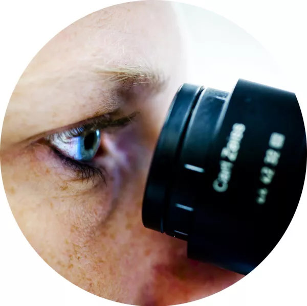
If you ever wondered how stem cells look like under a microscope, or at this very moment realised that you do, this workshop is for you! You will get a look at live blood stem cells, as well as mature blood cells, under the microscope.
Contact WS 3
Svetlana Soboleva, PhD student at the Division of Molecular Medicine and Gene Therapy
Hugo Åkerstand, PhD student at the Division of Molecular Hematology
Contact WS 4
Taha Sen, PhD student at the Division of Molecular Medicine and Gene Therapy
Abdul Ghani Alattar, PhD student at the Division of Molecular Medicine and Gene Therapy
5. The use of antibodies in stem cell research
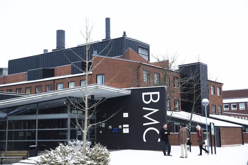
How do we distinguish a stem cell from a differentiated cell? How do we know that a protein is localized in the cytoplasm and not in the nucleus? In this workshop you will learn how antibodies work and how they are used in stem cell research. The workshop will start with a 15 minutes lecture, where we will talk about antibody structure and production, antigen-antibody interactions and their multiple applications. This will be followed by a hands-on activity at the BMC A12 microscope room where students will have the opportunity to witness antigen-antibody interactions live!
Contact
Hooi Min Tan Grahn, Postdoctoral fellow at the Division of Molecular Medicine and Gene Therapy
Sandra Capellera-Garcia, PhD student at the Division of Molecular Medicine and Gene Therapy
6 & 7. Behavioural assessment of neural stem cell transplants – From animal models to functional assessment
In this workshop we will present how researchers assess neural stem cells in vivo. We will give an introduction about research on laboratory animals, how we create models of neurodegenerative diseases, and how to assess the neural stem cells in vivo. The focus is on Parkinson’s disease and the transplantation of cells into the brain of rodents. After the workshop the student will have a basic idea about the animal models used for transplantation, how to assess graft survival and the immune-response, as well how a neural stem cell transplant can improve the behaviour of a rodent that has been rendered parkinsonian. The workshop will be based on presentations of the most commonly used behavioural test as well as some equipment. Due to the restricted nature of animal testing the practical part of the session will be limited.
Contact WS 6
Andreas Heuer, Visiting research fellow at Molecular Neurobiology
Contact WS 7
Marie Persson Vejgården, Biomedical analyst at Molecular Neurogenetics
Tiago Cardoso, PhD student at Developmental and Regenerative Neurobiology
Shelby Shrigley, PhD student at Developmental and Regenerative Neurobiology
8. Passera benmärgstromaceller
Benmärgstromacellerna skapar en essentiell miljö för blodstamcellerna i kroppen. Vi studerar därför de signaler som dessa stromaceller producerar för att kunna lära oss att expandera blodstamceller utanför kroppen. Detta skulle kunna öka chanserna för blodstamcellstransplantation. Stromacellerna växer snabbt utanför kroppen och måste därför passeras innan de blir överväxta. I denna workshop kommer man att få lära sig grunderna i cellodlingskonstens ABC - passera och räkna celler.
Contact
Mattias Magnusson, Ledare för forskargruppen Regulation of stem cells in normal and malignant hematopoiesis
Simon Hultmark, Doktorand i gruppen Regulation of stem cells in normal and malignant hematopoiesis
9 & 10. Snitta hjärnan och hitta transplantat
Ni kommer att få se hur man snittar en hjärna och hur man sen märker in den med speciella antikroppar för att kunna visualisera transplanterade stamceller. I mikroskopet letar vi sen upp cellerna och tittar på hur de transplanterade cellerna ser ut. Vi kommer även att visa hur man använder avancerad 3D imaging.
Kontakt WS 9
Malin Parmar, Ledare för forskargruppen Developmental and Regenerative Neurobiology
Ulla Jarl, Biomedicinsk analytiker i gruppen Developmental and Regenerative Neurobiology
Bengt Mattson, Neurobiology
Contact WS 10 (på engelska)
Daniel Tornero, Postdoctoral fellow at the Laboratory of Stem Cells and Restorative Neurology
Cecilia Laterza, Postdoctoral fellow at the Laboratory of Stem Cells and Restorative Neurology
11. Gene expression analysis: Are you faster than a robot?
In this workshop we will give an introduction on gene expression and how we can measure it by using qPCR. Moreover, we will introduce our pipetting robot to the students. They will then have the possibility of practicing pipetting and challenging our robot.
Contact
Per Ludvik Brattås, PhD student at Molecular Neurogenetics
Daniela Grassi, PhD student at Molecular Neurogenetics
12. Cord blood saves lives
Umbilical cord connects mother and her baby during pregnancy and after delivery can be used as a source to obtain stem cells. In this workshop we give you an overview on isolation of precious stem cells from cord blood samples. In theoretical part we will explain reasons to use blood stem cells in clinic and in research, with focus on cord blood derived stem cells. We will then move to the tissue culture lab and show you major steps of stem cell isolation from cord blood and how to assess the purity of isolated cells by flow cytometry.
Contact
Agatheeswaran Subramaniam, Postdoctoral fellow at the Division of Molecular Medicine and Gene Therapy
Shubhranshu Debnath, PhD student at the Division of Molecular Medicine and Gene Therapy
13. Can we measure how neurons function and communicate in the brain?
This workshop will give students a brief introduction on how you can assess the function of living cells, such as neurons from cell cultures or from tissue sections of animal models. How do neurons communicate with each other? Are there differences in the healthy or disease state? To answer these questions, we will apply whole-cell patch clamping, an electrophysiological measurement. After a short description of the technique will demonstrate it hands-on in the lab.
Contact
Daniella Ottosson, Postdoctoral fellow at Developmental and Regenerative Neurobiology
Marcella Birtele, PhD student at Developmental and Regenerative Neurobiology
14. Is the cell alive? Identify this using flow cytometry
We would like to explain to students the general ways to distinguish dead cells, viable cells or apoptotic cells with FACS analysis. Students will be asked to kill cells with different methods, such as applying heat-shock, detergent or ethanol on the cells. This hands-on activity will take about 15 min. The general principles will be explained in front of the instrument at BMC for another 15 min at B12 Canto room. Students will be encouraged to ask questions for another 10 min.
Contact
Anna Rydström, Research engineer at the Division of Molecular Medicine and Gene Therapy
Els Mansell, Postdoctoral fellow at the Division of Molecular Medicine and Gene Therapy
15. Cloning workshop: how to cut DNA and visualize it on a gel
In this workshop we will give an introduction on cloning and how it is used in the lab. The students will learn how to pipette a restriction enzyme reaction and how to visualize the cut plasmid DNA on an agarose gel.
Contact
Marie Jönsson, Postdoctoral fellow at Molecular Neurogenetics
Rebecca Petri, PhD student at Molecular Neurogenetics
16. Vem äger ditt DNA?
Är det ok att någon annan tjänar pengar på informationen som finns lagrad i dina egna celler? Och vem borde få veta vad ditt DNA kan avslöja om din framtid? I den här interaktiva workshopen kommer vi att diskutera olika fall kring vem som äger ditt DNA och testa vad vi tror/tycker är rätt och fel.
Contact
Sofie Singbrant Söderberg, Ledare för forskargruppen Stem cell and red cell biology
17. Building a human fetal brain in the dish with stem cells and bioengineering
In this workshop the students will hear about how to combine bioengineering and stem cell techniques to build brain-like structures from human stem cells. The workshop will involve demonstration of microfluidic techniques and looking at fluorescently labelled bioengineered tissue under the microscope.
Contact
Marc Isaksson, PhD student at Human Neural Development
Pia Wantiya, Student at Human Neural Development


