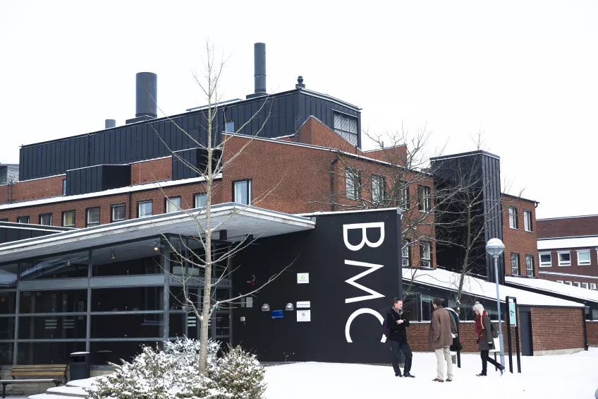
UniStem workshops 2018
Can you cut into DNA? How do blood stem cells look in the microscope? Can you decipher the code of life?
These and many more questions will be answered by motivated PhD students, postdoctoral fellows, and senior scientists during UniStem Day on March 16th, 2018.
Below you find a list of workshops and hands-on sessions that we will offer during UniStem Day at Lund Stem Cell Center. The workshops will be held in Swedish and/or English.
Welcome!
1. Direct neural reprogramming
This workshop will include a tour of the lab where cell culture is performed. In our lab, we reprogram skin cells from Parkinson’s disease patients into neurons. The students will be able to see the morphological changes that occur along the process in these cells, and get some insight about the procedures used for cell conversion (approximately 30 minutes) and perform a medium change. After that, they will have the chance to look at the cells at the stage where conversion is complete, under the fluorescence microscope (15 min).
Contact
Shelby Shrigley, PhD student at Developmental and Regenerative Neurobiology
Janelle Drouin-Ouellet, Postdoctoral fellow at Developmental and Regenerative Neurobiology
2. Cutting into DNA - genetic engineering approaches for science and medicine
Advanced overview of approaches and perspectives towards gene therapy and gene editing in science and medicine. The review will present the rational for gene correction and replacement strategies via viral and non-viral means, describe considerations for safety and efficacy in gene therapy approaches, and lastly, offer a glimpse into a potential design process for therapeutic gene editing approaches.
Contact
David Yudovich, PhD student at the Division of Molecular Medicine and Gene Therapy
Ludwig Schmiderer, PhD student at the Division of Molecular Medicine and Gene Therapy
3. Modelling brain diseases in a dish - how can we distinguish different cell types?
The workshop will start with a brief introduction (about 15 min) to the process of CNS disease modeling, and how one can use human pluripotent stem cells to generate human brain cell types, and how immunocytochemistry can help us identify them. Afterwards, the students will have a tour of our histology laboratory, where they will get a hands-on experience with cell staining (they will add DAPI to pre-stained cultures, to later visualize the cells under our inverted microscope (up to 30 min).
Contact
Carla Azevedo, PhD student at CNS Disease Modeling
Margarita Chumarina, PhD student at CNS Disease Modeling
4. Where to find stem cells in your body and how to use them to study and treat central nervous system pathologies
We will introduce the concept of what a stem cell is and where we can find sources of them in an adult. We will focus on bone marrow mesenchymal stromal cells and will explain the different approaches using them for treating pathologies affecting the central nervous system, with a special mention to brain tumour. Next, we will show the students our facilities, our lab and cell lab, where students will look at bone marrow stromal cells under the microscope.
Contact
Tania Ramos Moreno, Postdoctoral fellow at Glioma Cell Therapy group
Francesca Romana Stefani, PhD student at Glioma Cell Therapy group
5. Isolate and image cells - Flow cytometry, cell sorting and automated image analysis
Introducing students to the Flow Cytometry Sorting technique used to isolate cells obtaining pure populations of rare cell types. We will sort out fluorescently labeled cells into plates, to illustrate the difficulties and convenience of separating one cell type from all others using Fluorescence Activated Cell Sorting (FACS) (20 min). After that we will move to Cellomics High Content Screening and show students automated image analysis of cells. (20 min).
Contact
Teona Roschupkina, FACS Core Facility at Lund Stem Cell Center
Anna Hammarberg, MultiPark - Cellomics and Flow Cytometry Core Facility
6. The use of antibodies in stem cell research
How do we distinguish a stem cell from a differentiated cell? How do we know that a protein is localized in the cytoplasm and not in the nucleus? In this workshop you will learn how antibodies work and how they are used in stem cell research. The workshop will start with a 15 minutes lecture, where we will talk about antibody structure and production, antigen-antibody interactions and their multiple applications. This will be followed by a hands-on activity in the microscope room where students will have the opportunity to witness antigen-antibody interactions live.
Contact
Hooi Min Tan Grahn, Postdoctoral fellow at the Division of Molecular Medicine and Gene Therapy
Mayur Jain, Postdoctoral fellow at the Division of Molecular Medicine and Gene Therapy
8. Discover the machinery of a blood stem cell
Proteins are the building blocks that make up all the cells in your body as well as function as the cells’ machinery. In this workshop, you will discover proteins that are important for blood stem cells and how to study them. The workshop will start with a practical exercise in our lab where you will extract proteins from cells and determine their concentration. We will then give a short presentation where you will learn about a proteins composition and how it determines its properties, followed by a hands-on activity where you will use your new-found knowledge to practice methods commonly used in the study of proteins (proteomics).
Contact
Kristýna Pimková, Postdoctoral fellow at Proteomic Hematology
Maria Jassinskaja, PhD student at Proteomic Hematology
9. How to separate cancer stem cells from normal stem cells using single-cell technology
In this workshop we will introduce the single-cell qPCR technology where each cell's gene expression is measured and used to separate healthy and cancerous stem cells. The students will learn to pipette in small chips and load the sample in the machine.They will also look at results to see the molecular differences between each single cancer stem cell and a normal cell.
Contact
Mikael Sommarin, PhD student at the Division of Molecular Hematology
Fatemeh Safi, PhD student at the Division of Molecular Hematology
10 & 11. Blood stem cells under the microscope
If you ever wondered how stem cells look like under a microscope, or at this very moment realised that you do, this workshop is for you! You will get a look at live blood stem cells, as well as mature blood cells under the microscope.
Contact WS 10
Svetlana Soboleva, PhD student at the Division of Molecular Medicine and Gene Therapy
Hugo Åkerstand, PhD student at the Division of Molecular Hematology
Contact WS 11
Abdul Ghani Alattar, PhD student at the Division of Molecular Medicine and Gene Therapy
Alban Johansson, Master student at the Division of Molecular Medicine and Gene Therapy
12. Exploring the cell diversity of the brain
Do you know that neurons are not the only cell type in the human brain? If you are wondering what cells you can find in the brain, join this workshop! We will tell you about the cells that form the brain and how these cells look like under the microscope. Thereafter, you will also have the opportunity to take care of them.
Contact
Ana Gonzalez Ramos, PhD student at Experimental Epilepsy
Eliška Waloschková, PhD student at Experimental Epilepsy
13. Behavioural assessment of neural stem cell transplants - From animal models to functional assessment
In this workshop we will present how researchers assess neural stem cells in vivo. We will give an introduction about research on laboratory animals, how we create models of neurodegenerative diseases, and how to assess the neural stem cells in vivo. The focus is on Parkinson’s disease and the transplantation of cells into the brain of rodents. After the workshop the students will have a basic idea about the animal models used for transplantation, how to assess graft survival and the immune-response, and also how a neural stem cell transplant can improve the behaviour of a rodent that has been rendered parkinsonian. The workshop will be based on presentations of the most commonly used behavioural test as well as some equipment. Due to the restricted nature of animal testing the practical part of the session will be limited.
Contact
Tiago Cardoso, PhD student at Developmental and Regenerative Neurobiology
Deirdre Hoban, Postdoctoral fellow at Developmental and Regenerative Neurobiology
Andrew Adler, Postdoctoral fellow at Developmental and Regenerative Neurobiology
14. Snitta hjärnan och hitta transplantat
Ni kommer att få se hur man snittar en hjärna och hur man sen märker in den med speciella antikroppar för att kunna visualisera transplanterade stamceller. I mikroskopet letar vi sen upp cellerna och tittar på hur de transplanterade cellerna ser ut. Vi kommer även att visa hur man använder avancerad 3D imaging.
Kontakt
Ulla Jarl, Biomedicinsk analytiker i gruppen Developmental and Regenerative Neurobiology
Bengt Mattson, Developmental and Regenerative Neurobiology
Olof Torper, Postdoctoral fellow at Developmental and Regenerative Neurobiology
15. Deciphering the code of life
Your identity, what disorders you could get and how your brain creates thoughts is directed by your DNA, a string of more than 3 billion nucleotides residing inside your cells. How do scientist make sense of such a complex code? In this workshop we will demonstrate how mathematics and computer science will be key to understanding human disorders, as well as comprehending life itself.
Contact
Per Ludvik Brattås, PhD student at Molecular Neurogenetics
Daniela Grassi, PhD student at Molecular Neurogenetics
16. Transplant cells into the brain and find them
Do you ever wonder how scientist are able to study human cells into the mouse brain? In this workshop students will learn and practice how to transplant cells (mock injections) inside the brain (gelatine brains), they will practice the process of cutting a brain in thin slices, mark the proteins of different cell types using specific antibodies that recognise them and mount the sliced sections on slides to visualise them under the microscope. For each step, students will first get a short introduction and later they will be able to practice the techniques. They will be encouraged to ask questions along the workshop.
Contact
Jonas Fritze, PhD student at Stem Cells, Aging and Neurodegeneration
Ella Quista, PhD student at Stem Cells, Aging and Neurodegeneration
Isaac Canals, Postdoctoral fellow at Stem Cells, Aging and Neurodegeneration
Sara Palma, Postdoctoral fellow at Laboratory of Stem Cells and Restorative Neurology
17. From DNA to proteins - an unexpected journey
Would you like to know how the information from your DNA is converted into the building blocks of the cell? During this workshop we will describe in a short lecture (20 min) the journey that takes DNA to become a protein and which methods we use in the lab to visualize proteins. Then you will have the opportunity to try yourself some of these techniques in a practical session (20 min).
Contact
Giulia Beneventi, PhD student at the Division of Molecular Hematology
Nicola Guzzi, PhD student at the Division of Molecular Hematology
18. Counting cells like a pro!
This workshops aims to give a brief introduction into cell counting and its basic principles. We will then move to the culture lab to demonstrate manual cell counting of human primary mesenchymal stem cells with a traditional hemocytometer and parallel automated cell counting on a NC250 machine.
Contact
Dimitra Zacharaki, PhD student at the Division of Molecular Hematology
Franziska Olm, PhD student at the Division of Molecular Hematology
Sandro Meyer, PhD student at the Division of Molecular Hematology